


Persistent pain in the pelvic area in women is often associated with circulatory problems in this area. Latent dilation of the pelvic veins is not uncommon. What it is and how to treat it - we will examine in this material!
As shown by medical statistics, more than half of middle-aged women experience recurrent lower abdominal pain. About half of all these cases are associated with circulatory disorders. This is manifested by blood stagnation and subsequent shedding of intercellular fluid into the pelvic cavity. Closure leads to compression of the soft tissues of the organs. This provokes the development of pain syndrome. The cause of this pathological process are varicose veins of the pelvis.
It usually begins to develop during pregnancy and then progresses slowly throughout a woman's life. Currently, there is no reliable data on the causes of this phenomenon and on effective treatment methods.
Development mechanism
In a normally functioning vein, blood flows only in one direction. The backflow is prevented by the valve system. In case the valves lose their integrity and elasticity, a gradual reverse venous blood flow develops. With a prolonged pathological process, this leads to constant blood stagnation. As a result, the vascular wall stretches and the vein cavity expands. It loses bandwidth and the ability to compress when needed.
In the initial stage, the pain in this disease appears due to damage to the nerve endings innervating the vascular walls of the venous bed.
Possible causes
Currently, science does not know the exact cause of this disease. Possible risk factors include the following.
- Physiology of pregnancy. During pregnancy, a significant increase in circulating blood volume is observed. This leads to an increase in the weight of the pregnant woman. It is believed that excess blood volume in combination with excess body weight contributes to the expansion of the venous bed. In the future, this provokes overload and damage to the venous valves.
- The action of estrogens. During pregnancy, large doses of the hormone estrogen are constantly thrown into a woman's body. They are necessary for the preservation and growth of the fetus. Estrogens reduce the risk of miscarriage by relaxing the muscles in the uterus. But on the other hand, these substances negatively affect the contractility of blood vessels.
- Individual anatomical disorders. In some patients individual anatomical features are revealed in relation to the pelvic veins. Their location is, in principle, unfavorable for the onset of pregnancy. Therefore, the onset of fertilization in most cases leads to the development of venous insufficiency.
Is there a link between this condition and varicose veins of the lower extremities?
Varicose veins in the small pelvis are very similar to the condition of varicose veins in the legs. In both cases, the valves in the veins that help the blood flow to the heart are affected. The function of the valves to prevent blood flow is impaired. When the valves collapse, blood gets stuck in the veins. The filling veins stretch and worsen the blockage. Pelvic venous overload syndrome develops mainly near the uterus, fallopian tubes, vulva and even the vagina. The condition is usually accompanied by weight gain, which is inevitable during pregnancy.
Varicose veins are commonly seen in women:
- from 20-45 years old;
- during multiple pregnancies.
What are the signs and symptoms?
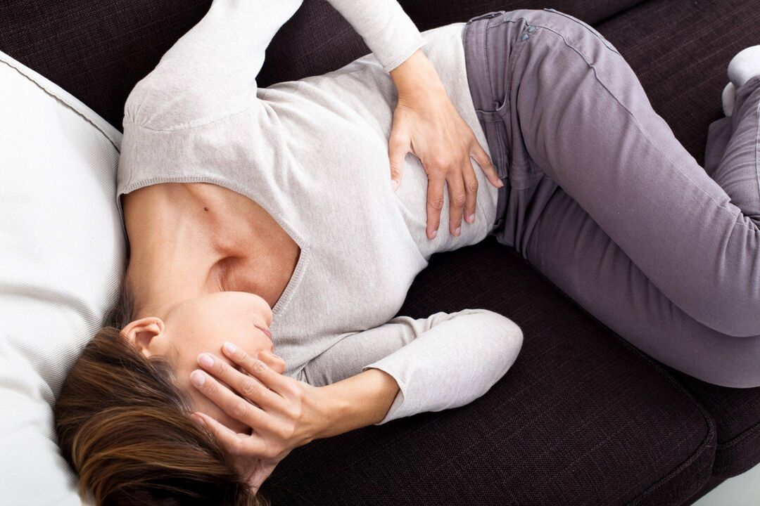
The most common complaint of an injured woman is pain of varying severity. The pain syndrome is constant in nature and not cyclical in nature. Increased pain appears:
- before the onset of menstruation;
- at the end of a hard day at work;
- after standing for a long time;
- during or immediately after sexual intercourse;
- in the later stages of pregnancy.
All of these symptoms are reason enough to see a phlebologist. This condition can be accompanied by a periodic increase in total body weight by 2-5 kg. This weight is formed mainly due to the shedding of fluid in the abdominal cavity of the small pelvis.
There are many other non-specific symptoms that appear with varying intensity. In general, the symptoms are more likely to appear at the end of the day or after prolonged standing or even after sexual intercourse. In some cases, the pain can be severe and affect personal and social relationships.
Signs may also include:
- swelling of the vulva and vagina;
- varicose veins of the external genitalia, buttocks, legs;
- abnormal menstrual bleeding;
- pain when touching the lower abdomen;
- pain during intercourse;
- painful periods;
- backache;
- vaginal discharge;
- general weakness and apathy;
- feelings of depression and depression.
In most cases, the presence of pelvic stasis syndrome is not obvious and the diagnosis can only be made after other diseases have been ruled out. Similar disorders that may have the same symptoms include:
- endometriosis;
- uterine fibroids;
- uterine prolapse (the uterus sinks lower into the pelvis as a result of weak pelvic floor muscles).
Diagnosis and laboratory research
For a complete diagnosis of the presence of stagnation, laboratory tests are important. A woman is usually assigned a standard set of examinations.
Ultrasound examination of the pelvic organs. It will help assess the condition of the uterus and other organs of the small pelvis. It can also help visualize blood flow and the presence of varicose veins in the pelvis. The procedure is painless and takes about 30 minutes. Generally cheap and effective.
Phlebograms. This test has been widely used in the past to diagnose blood stagnation in the pelvic cavity, but today, if possible, the procedure is replaced by computed tomography. The test involves injecting a special dye into a vein in the groin and then using X-rays. The procedure takes about 30-45 minutes and is performed on an outpatient basis. Examination is painless, however, there is a risk of developing an allergic reaction to the contrast agent. Also, the possibility of exposure to radiation of the pelvic organs is not excluded.
Computed tomography is often used in the diagnosis of pelvic varicose veins. This method allows you to visually examine the anatomy of the pelvis and identify varicose veins of the pelvis. This is due to radiation exposure and is not recommended as a test for pregnant women.
Magnetic resonance imaging is a very useful test in diagnosing pelvic congestion syndrome. Does not use radiation and contrast agent. This is a painless examination. The images are of excellent quality. It is the preferred method of choice for diagnosing most cases. The test lasts about 15 minutes and is performed on an outpatient basis.